Technology is developing fast every single day, and because of it, we have better and safer lives. One of the biggest advancements in technology is noticed in medicine, and there are a lot of different screenings that are faster, cheaper, and safer just because of new machines and computers. Some of the most commonly used examinations include the CT scan and the MRI, and here we are going to tell you a bit more about these two screenings and what are they used for.
Table of Contents
What are these examinations and what are they used for?
Computerized tomography examinations work in the way that they take images of the body from different angles using X-rays. After the images of the body are taken, the machine combines them and creates the final result that the specialist can read and understand. This procedure is fast, cheap, and efficient and it is used to examine everything starting from your bone structure, soft tissues, and all the internal organs you have.
Computerized tomography is most commonly used to determine lung issues, head conditions, bone structure problems, and circulation issues.
On the other hand, magnetic resonance imaging combines the technology of radio waves and magnetic fields to capture the image of your internal organs. With it, some of the bone structure can be shown, but it is mostly used to examine the organs, and the soft tissues surrounding them. MRI scans are said to be more effective when it comes to detailed imaging, but they are also more expensive and they come with higher risks.
MRI is most commonly used to determine breast cancers, tumors, liver disease, brain conditions, and joint abnormalities.
Now let’s talk about the main differences between these two examinations, what are the benefits from them, and what are the possible risks you should be aware of before deciding to get the scan.
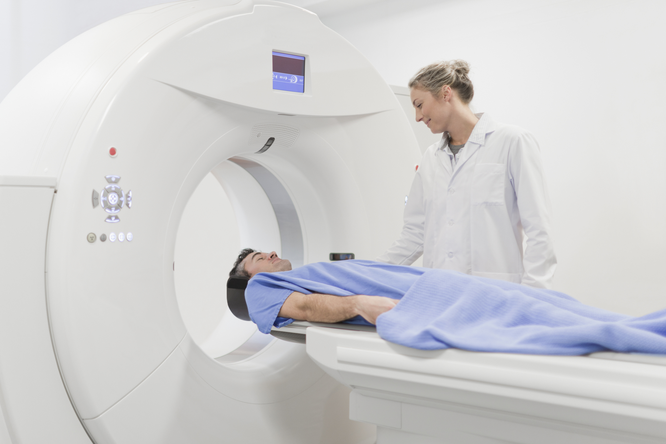
img source: bodygears.com
Main Differences
Even though these scans are used to show the condition of your body and internal organs, they still have some differences in them. The benefits and the risks are some of the things that show the differences better, and we will go into detail about that.
In short, magnetic resonance imaging is usually more expensive than a computerized tomography, but it also shows better images and more details. The CT screenings are widely used, and in most cases, GPs usually recommend them first because the whole process is faster, easier, and cheaper.Read more about CT scan in detail here on LabsAdvisor.com.
The biggest difference between the two is that computerized tomography is done with X-rays, and MRI uses a different technology. Now let’s talk about the benefits as well as the risks that come with these screenings.
Risks

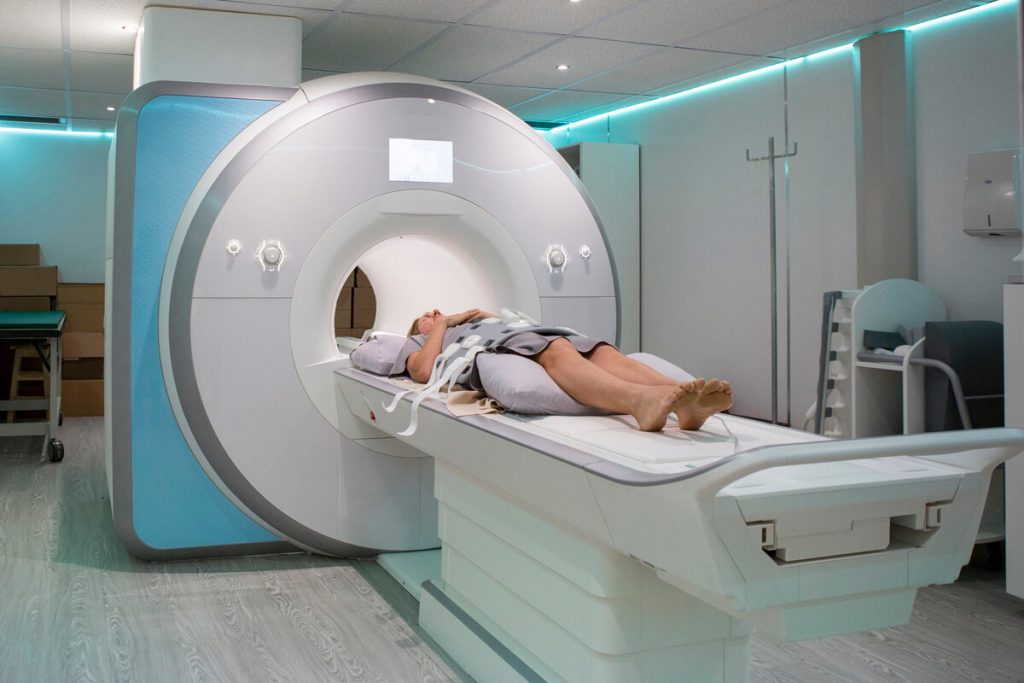
img source: scientist.com
There are risks that come with both scans, and because of that, you need to consult with your GP or a specialist about which type would not be harmful to you.
When it comes to CT, one thing that should always be paid attention to is pregnant women. This type of scan can be really dangerous to the fetus and it may harm them. Since this procedure uses X-rays, there is a dose of radiation that the patient is exposed to. Because of this, you should not get this type of scan too frequently. Last, but not least, some people may exhibit allergic reactions to the dye that is used during the examination.
On the other hand, MRI may be harmful to people who have undergone previous procedures. In case you are suffering from heart disease and if you have a pacemaker, this is not the right type of examination for you. If you have an intrauterine device or artificial joints, you have to notify your GP about these things.
The machine is loud and it is also a smaller, confined space. If you suffer from anxiety, panic attacks, or claustrophobia, this may be a negative experience for you. Some people have reported having an allergic reaction to the magnets, and loud noises may lead to hearing problems.
Benefits
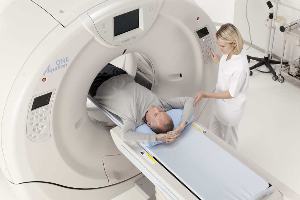
img source: dicardiology.com
The main benefit of both types of examinations is that the specialist will be able to see if there is anything wrong in your body if you are suffering from a disease, how fast it is progressing, and at what stage it is.
Magnetic resonance imaging is commonly used to find out if there is an abnormal tissue in your organs, and this scan gives a better-quality image. On the other hand, computerized tomography is more commonly used and it is a faster type of examination. With it, your GP will be able to see the internal organs, your skeleton as well as the tissues surrounding them.
Depending on the type of issue you are being examined for, your specialist can recommend one of the two, and according to wispecialists.com, they may even suggest an additional ultrasound or dual-energy x-ray scans.
Exploring Alternative Imaging Modalities
While CT and MRI scans are prominent in medical imaging, exploring alternative modalities like ultrasound and PET scans unveils a broader spectrum of diagnostic tools, each with unique advantages tailored to specific medical conditions. For a diverse experience in medical scans you should first visit this website.
Ultrasound: Clarity in Real-Time
Ultrasound imaging, utilizing high-frequency sound waves, offers real-time insights into the body’s inner workings. Its non-invasive nature and absence of ionizing radiation make it a preferred choice for monitoring fetal development during pregnancy. Moreover, its excellence in visualizing soft tissues and fluid-filled structures positions it as a go-to option for evaluating organ conditions, particularly in the abdomen and pelvis.
PET Scans: Unveiling Metabolic Insights
PET scans, on the other hand, excel in detecting metabolic changes at a cellular level, often before structural changes become apparent on CT or MRI. By introducing a radioactive tracer into the body, PET scans illuminate abnormal metabolic activity, invaluable in oncology for identifying cancerous growths and assessing treatment effectiveness. Additionally, its applications extend to neurology, where it aids in diagnosing conditions like Alzheimer’s by highlighting brain function anomalies.
Each imaging modality shines under different circumstances, and understanding their distinct advantages ensures a more comprehensive approach to patient diagnosis and treatment. As medical technology advances, embracing this diversity in imaging techniques not only enriches diagnostic accuracy but also paves the way for personalized treatment strategies.
How to choose what you need?

img source: ucsfhealth.org
Your doctor will give you their professional opinion based on your specific case and diagnosis. You should never try and get these scans on your own because even though they are safe, you should not expose your body to the rays too often.
A specialist is needed later on to see the results you are going to get, and they will you more about the things you are dealing with. However, in case you need to know more about the condition of your joints, internal tissues and organs, chances are you are going to need magnetic resonance imaging. On the other hand, if the doctors need to know more about the condition of your internal organs after an accident that caused damage to your head or bones, you are more likely to need a computerized tomography.
As you can see, these screenings are crucial for early diagnosis, and they can help to determine the course of therapy for the patient. You should know that even though there are some risks that come with these examinations, the screenings are safe. You should always go for regular checkups and talk to your GP about hereditary diseases and what you can do to catch them in the early stages.
Thanks to these screenings, we are able to have better lives, cope with the symptoms of serious conditions and we are able to prolong our life. Never choose the screening on your own without consulting a specialist, and always choose a well-renounced clinic and imaging specialists.